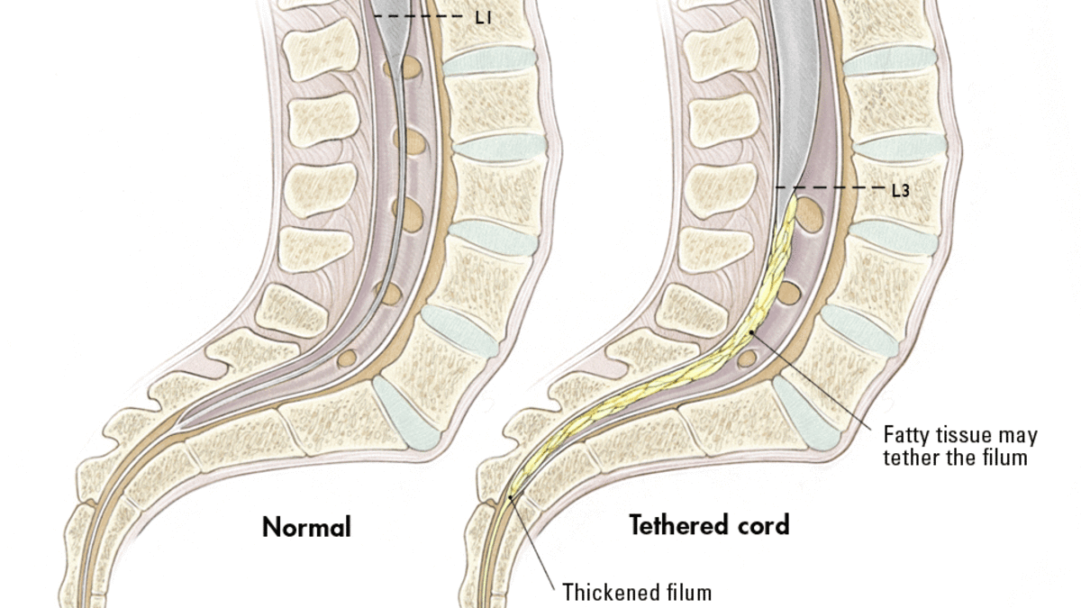
Tethered Cord Syndrome
OVERVIEW
Tethered spinal cord syndrome is a condition where the spinal cord is stuck or “tethered” to the tissues around it, instead of moving freely inside the spine. This happens because of problems very early in development, such as failure of dysjunction (when the spinal cord doesn’t properly separate from the skin) or failure of retrogressive differentiation (when the lower end of the spinal cord does not regress normally). This pulling creates tension on the spinal cord, which over time can cause stretching, reduced blood flow, and nerve problems such as pain or weakness.

It often happens in children born with spina bifida occulta. In this condition, incomplete closure of the neural tube leads to small hidden defects beneath the skin. When retrogressive differentiation fails, the lower portion of the embryonic spinal cord does not regress as it should, leaving behind an abnormally thickened or fatty filum terminale—a band of tissue that can tether the cord in a lower-than-normal position. Normally, the filum terminale is a thin, flexible strand that simply anchors the cord, but when it is abnormally formed, it acts like a tight band that restricts movement. In some cases, fatty tissue outside the spinal canal may also connect to the cord, further limiting its mobility.
Even though the skin looks closed at birth, the spinal cord may still be tethered underneath. As a child grows, the cord gets stretched tighter, which can lead to neurological problems and symptoms as they get older.
Filum terminale and tethered cord pathology remain underrecognized in the context of rare diseases, yet there is growing evidence that they may share developmental origins with certain genetic syndromes. For example, associations with VACTERL syndrome have been described, likely reflecting a common dysfunction in embryonic lineage development that disrupts both neural tube closure and mesodermal differentiation. These overlaps highlight the need for greater awareness of tethered cord manifestations in patients with syndromic presentations, as subtle filum abnormalities may contribute to neurological morbidity but remain overlooked.
SYMPTOMS & TYPES
In addition to myelomeningocele and lipomyelomeningocele discussed above, the following are other causes of tethered cord that vary in severity of symptoms and treatment:
-
Dermal sinus tract (a rare congenital deformity)
-
Diastematomyelia (split spinal cord)
-
Lipoma (a benign, fatty growth)
-
Tumor
-
Thickened/tight filum terminale (a delicate filament near the tailbone)
-
A history of spine trauma
-
A history of spine surgery
Symptoms in Children
The symptoms listed below are some of the ways in which tethered spinal cord syndrome may be exhibited in children:
-
Lesion on the lower back
-
Fatty tumor or deep dimple on the lower back
-
Skin discoloration on the lower back
-
Hairy patch on the lower back
-
Back pain, worsened by activity and relieved with rest
-
Leg pain and leg aches, especially as a result of activity
-
Leg numbness or tingling
-
Changes in leg strength
-
Deterioration in gait
-
Progressive or repeated muscle contractions
-
Leg deformities
-
Spine tenderness
-
Scoliosis (curvature of the spine)
-
Bowel and bladder problems
-
Walking on tip-toes
A patient with tethered spinal cord can continue undiagnosed into adulthood. In such cases, the strain on the spinal cord increases, leading to increasing sensory and motor problems, as well as loss of bladder and bowel control.
TESTING & DIAGNOSIS
If a tethered cord is suspected, one or more tests may be necessary to confirm the diagnosis.
-
MRI: A diagnostic test that, using powerful magnets and computer technology, produces three-dimensional images of body structures; can show the spinal cord, nerve roots, and surrounding areas, as well as enlargement, degeneration, and displacement
-
Myleogram: An x-ray of the spinal canal following injection of a contrast material into the thecal sac; can show pressure on the spinal cord or nerves caused by tethered spinal cord
-
CT or CAT scan: A diagnostic image created after a computer reads x-rays; may be used after a myelogram to show how the dye flows around the spinal cord and nerves
-
Ultrasound: A water-soluble gel is placed on the skin where the transducer (a handheld device that directs the high-frequency sound waves to the spine) is to be placed. The gel helps to transmit the sound to the skin surface. The ultrasound is turned on, and images of the spinal cord moving in the thecal sac are obtained. This is a technique that can only be used in the first months of life while the spine itself is not yet mature.
It's important to know that tethered cord syndrome is often occult, meaning it may not show up clearly on imaging like MRI or ultrasound. In many cases, diagnosis depends on recognizing clinical symptoms and changes over time, rather than relying solely on scans.
TREATMENT & CARE
Surgery
Microsurgical resection of the filum terminale is a surgery used to release a tethered spinal cord. A small opening is made in the lower back, and with the help of a microscope, the surgeon carefully finds the filum terminale—a small band of tissue that is abnormally thick or fatty. Once identified, this band is safely cut, freeing the spinal cord so it can move more normally. The goal is to relieve tension on the cord, prevent further damage, and sometimes improve symptoms such as pain or weakness.
The combined complication rate of this surgery is usually less than 5%. Complications include infection, cerebrospinal fluid leak and temporary urinary retention or leg paraesthesias. There is a risk of retethering, because symptoms of tethering can occur during periods of growth, 5 to 7% of children with this syndrome require repeated surgery.
Followup
Treatment of a tethered cord syndrome does require follow-up with the treating neurosurgeon and typically with physical therapy/ occupational therapy. After some time, follow-up with the neurosurgeon may be on an as needed basis or if symptoms return.
SOURCES
The information on this page is sourced from the American Association of Neurological Surgeons (AANS), a trusted resource for neurosurgical education and patient information.